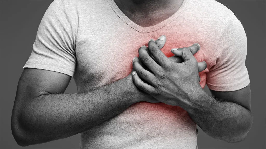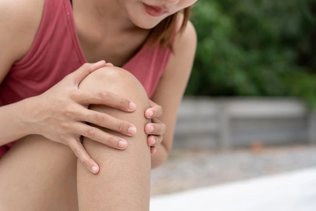
Knee pain is a prevalent health issue that can significantly impact the quality of life, particularly in females. Whether you’re an active individual or experiencing discomfort during daily tasks, knee pain can stem from a variety of causes. Understanding the underlying reasons for knee pain in females is crucial for effective management and treatment. In this comprehensive guide, we will explore the various factors that contribute to knee pain in women, common symptoms to watch for, and the treatment options available to help alleviate the discomfort
Understanding Knee Pain in Females
Knee pain in females is not a one-size-fits-all condition. Women experience knee pain for several reasons, ranging from age-related wear and tear to injuries caused by physical activity. Additionally, factors such as hormonal fluctuations, anatomical differences, and lifestyle choices can contribute to knee discomfort. The knee joint is one of the most complex and largest joints in the body, so pain in this area can significantly affect mobility and overall function.
Hormonal Differences and Knee Pain in Women
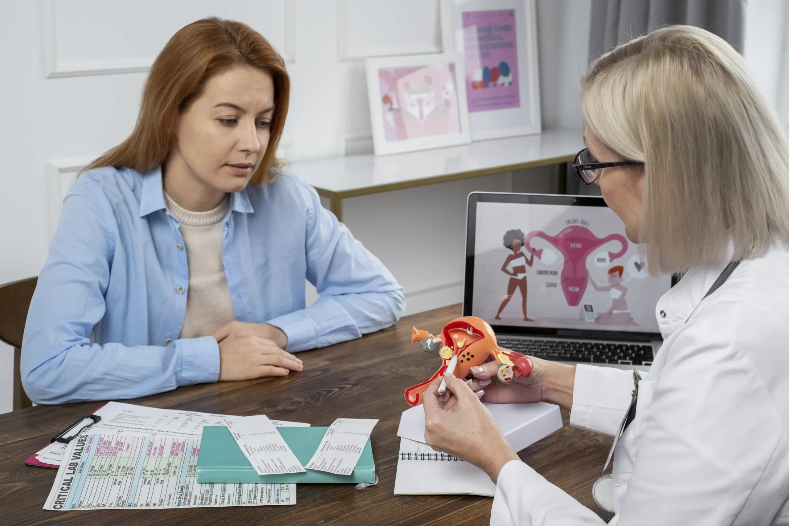
One of the primary causes of knee pain in females is hormonal fluctuations, particularly those associated with menstruation, pregnancy, and menopause. Studies have shown that women are more likely to develop knee pain than men due to the hormonal differences that impact ligaments and tendons.
During menstruation, increased levels of the hormone relaxin can cause the ligaments to become looser, which may lead to joint instability and discomfort. During pregnancy, the body undergoes similar hormonal changes that can result in an increased risk of knee pain due to added weight and pressure on the joints. In post-menopausal women, decreased estrogen levels can affect bone density and joint health, leading to conditions like osteoarthritis.
Common Causes of Knee Pain in Females
1. Osteoarthritis (OA)
Osteoarthritis is one of the most common causes of knee pain in both men and women. However, it is particularly prevalent among women over the age of 50. OA occurs when the cartilage that cushions the knee joint deteriorates over time, leading to pain, swelling, and stiffness. This condition is often aggravated by physical activity and may worsen with age. In females, the risk of OA increases after menopause due to changes in estrogen levels, which are thought to play a role in maintaining joint health.
Symptoms of Osteoarthritis:
- Pain during or after activity
- Stiffness in the knee, particularly after sitting for extended periods
- Swelling and inflammation
- A feeling of instability or weakness in the knee
2. Patellofemoral Pain Syndrome (PFPS)
Patellofemoral Pain Syndrome, commonly known as “runner’s knee,” is another condition that often affects females. It is characterized by pain around the kneecap, especially when climbing stairs, squatting, or sitting for long periods. PFPS is more common in women due to differences in anatomy, such as wider hips and altered biomechanics. These factors can affect the alignment and tracking of the patella (kneecap), leading to pain and discomfort.
Symptoms of PFPS:
- Pain around or behind the kneecap
- Pain during activities like squatting, climbing stairs, or sitting for long periods
- Tenderness around the patella
3. Ligament Injuries (ACL Tears)
Anterior cruciate ligament (ACL) tears are common injuries, especially among active females involved in sports. The ACL is one of the primary ligaments in the knee, and when it is torn, it can lead to severe pain, instability, and swelling. Female athletes are more likely to suffer ACL injuries than their male counterparts, partly due to differences in muscle strength, joint stability, and hormonal influences.
Symptoms of an ACL Tear:
- Sudden, severe knee pain
- Swelling and bruising
- A feeling of instability or the knee “giving way”
- Difficulty bearing weight on the affected leg
4. Meniscus Tears
The meniscus is a piece of cartilage that acts as a cushion between the bones in the knee joint. Meniscus tears can occur due to aging, wear and tear, or traumatic injuries such as twisting the knee during sports activities. Women, especially those over the age of 40, are more prone to degenerative meniscus tears, which develop gradually over time.
Symptoms of Meniscus Tears:
- Knee pain, especially during twisting or turning motions
- Swelling or stiffness in the knee
- A popping sensation at the time of injury
- Limited range of motion or difficulty straightening the leg
5. Bursitis
Bursitis is the inflammation of the bursae, small fluid-filled sacs that reduce friction and cushion pressure points between the bones and tendons. When these sacs become inflamed, typically due to repetitive movement or pressure, bursitis can cause pain and swelling in the knee. This condition is more common in women who engage in activities such as running, cycling, or kneeling.
Symptoms of Bursitis:
- Swelling around the knee joint
- Tenderness or pain when touching the affected area
- Pain that worsens with activity or pressure on the knee
6. Tendonitis
Tendonitis refers to inflammation of the tendons, which are the fibrous tissues that connect muscles to bones. In the knee, tendonitis often affects the patellar tendon, which connects the kneecap to the shinbone. Overuse or repetitive strain from physical activities such as jumping, running, or squatting can lead to tendonitis. Women, especially those involved in sports, are susceptible to this condition.
Symptoms of Tendonitis:
- Pain and tenderness at the front of the knee, near the patella
- Swelling around the affected tendon
- Pain that worsens with activity, especially when jumping or running
7. Gout
Gout is a type of inflammatory arthritis that occurs when uric acid builds up in the bloodstream, forming crystals that deposit in the joints. Although it most commonly affects the big toe, gout can also cause knee pain in females. Gout attacks are often sudden and severe, with intense pain, swelling, and redness in the affected joint.
Symptoms of Gout:
- Intense, sudden pain in the knee
- Swelling and redness around the knee joint
- Warmth in the affected area
Risk Factors for Knee Pain in Females
Several factors can increase the likelihood of developing knee pain in females, including:
- Age: As women age, the risk of developing conditions such as osteoarthritis or meniscus tears increases.
- Hormonal Changes: Fluctuating hormones, especially during pregnancy, menopause, and menstruation, can affect joint stability.
- Activity Level: High-impact sports or physical activities that put stress on the knee joint can increase the risk of injuries and pain.
- Obesity: Excess body weight places additional strain on the knees, contributing to conditions like osteoarthritis.
- Injury History: Previous knee injuries, such as ACL tears or meniscus damage, can predispose females to future knee problems.
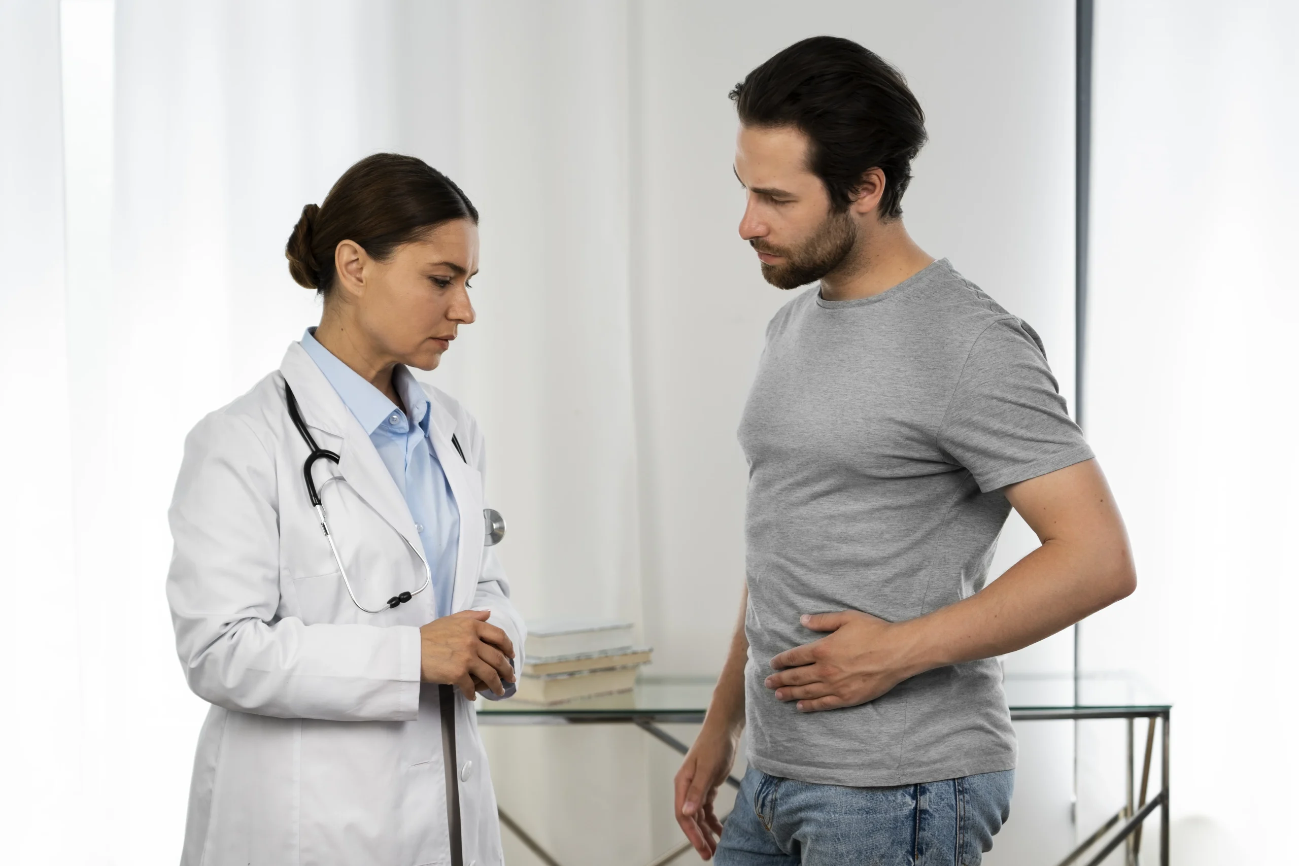
Diagnosis of Knee Pain in Females
If you’re experiencing knee pain, it’s important to seek medical attention for an accurate diagnosis. Your healthcare provider will likely perform a physical examination, ask about your symptoms and activity level, and may recommend diagnostic tests such as:
- X-rays to check for bone abnormalities or signs of arthritis
- MRI scans to assess soft tissue damage, such as ligament or tendon injuries
- Ultrasound to detect inflammation in the bursae or tendons
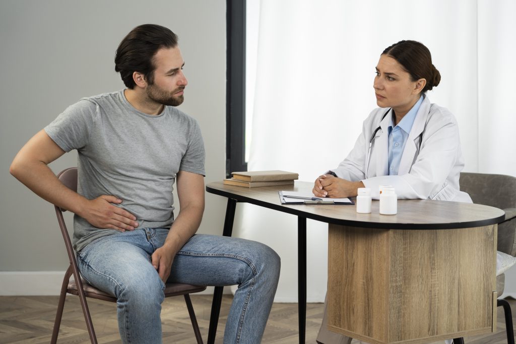
Treatment Options for Knee Pain in Females
The treatment for knee pain depends on the underlying cause and severity of the condition. Common treatment options include:
1. Conservative Treatments
- Rest: Taking time off from physical activities can allow the knee to heal and reduce inflammation.
- Ice and Heat Therapy: Applying ice packs or heating pads can help reduce swelling and alleviate pain.
- Physical Therapy: Strengthening the muscles around the knee can improve joint stability and reduce pain.
- Medications: Non-steroidal anti-inflammatory drugs (NSAIDs) such as ibuprofen can reduce pain and inflammation.
2. Surgical Treatments
In some cases, surgical intervention may be necessary, especially if conservative treatments fail. Surgical options may include:
- Arthroscopy: A minimally invasive procedure to remove damaged tissue or repair ligaments.
- Knee Replacement Surgery: In cases of severe osteoarthritis or joint degeneration, knee replacement may be recommended to relieve pain and restore function.
Prevention of Knee Pain in Females
While some causes of knee pain cannot be entirely prevented, there are steps you can take to reduce your risk:
- Maintain a healthy weight to minimize stress on the knees
- Perform strengthening exercises for the quadriceps and hamstrings to support the knee joint
- Avoid high-impact activities or modify your exercise routine if you’re prone to injuries
- Wear supportive footwear, especially when participating in physical activities
Conclusion
Knee pain in females can arise from a variety of causes, ranging from hormonal changes to overuse injuries and degenerative conditions like osteoarthritis. By understanding the underlying factors and symptoms, women can take steps to manage and treat knee pain effectively. If you’re experiencing persistent knee pain, it’s essential to consult with a healthcare provider who can offer a personalized treatment plan.
At Kolekar Hospital, our team of orthopedic specialists is dedicated to providing the latest and most effective treatment options for knee pain. Whether you need physical therapy, medication, or advanced surgical solutions, we’re here to help you return to a pain-free life. Our goal is to help you regain mobility, improve your quality of life, and reduce pain through personalized care plans tailored to your unique needs.
For more information or to book an appointment, contact Kolekar Hospital today. Your health and comfort are our top priority, and we’re here to help you every step of the way.
![]()