Ignoring appendix symptoms can be risky because appendix removal surgery is often the safest treatment once appendicitis is suspected. What starts as mild stomach pain can quickly worsen and lead to serious complications if delayed. Doctors recommend timely appendix removal surgery to prevent rupture, infection, and prolonged recovery.
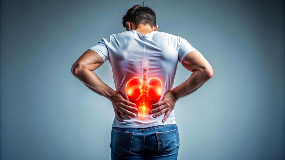
Why early attention matters
-
Appendix inflammation can worsen within hours
-
Risk of rupture increases if treatment is delayed
-
Early surgery usually means faster recovery and fewer complications
Key Conditions Treated
This service focuses on diagnosing and treating appendix-related conditions that require careful medical evaluation.
Common conditions include:
-
Acute appendicitis
-
Recurrent or chronic appendicitis
-
Inflamed or infected appendix
-
Complicated or ruptured appendix cases
Early identification of these conditions helps doctors decide the right time for surgery.
Diagnostic Services
Proper diagnosis is essential before recommending appendix removal surgery. Doctors use a combination of clinical assessment and tests to confirm the condition.
Diagnostic methods commonly used:
-
Physical examination to assess pain location and severity
-
Blood tests to detect infection or inflammation
-
Ultrasound or CT scan to confirm appendicitis and rule out other causes
Accurate diagnosis ensures safe and timely treatment.
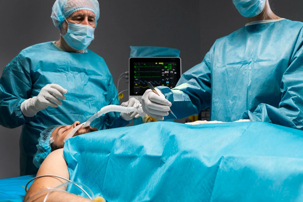
Treatment Options
Appendix removal surgery, also known as appendectomy, is the most effective treatment for appendicitis.
Surgical options may include:
-
Laparoscopic appendectomy: Minimally invasive surgery with smaller cuts, less pain, and quicker recovery
-
Open appendectomy: Recommended in complicated or ruptured appendix cases
The choice of surgery depends on the patient’s condition and overall health.
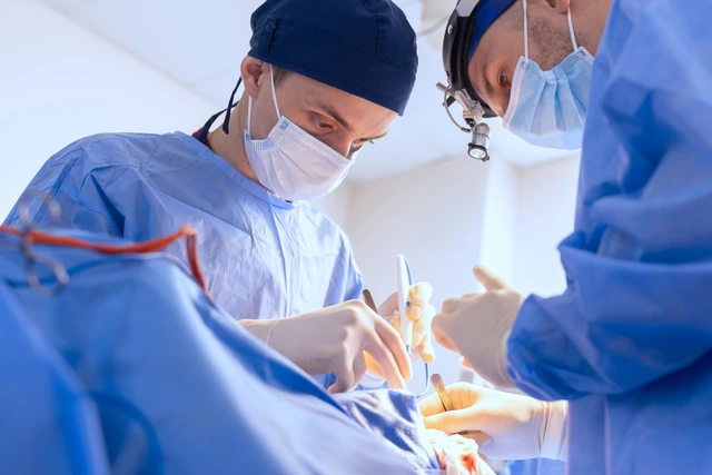
Why Choose Kolekar Hospital
Our hospital follows a patient-first and safety-focused approach to appendix care.
What patients can expect:
-
Experienced doctors and surgical team
-
Modern operation theatres and diagnostic facilities
-
Clear explanation of condition and treatment plan
-
Supportive care before and after surgery
The goal is safe treatment with comfort and confidence
FAQ
Pain that worsens, shifts to the lower right abdomen, or comes with fever, nausea, or vomiting should be evaluated immediately.
In most confirmed cases of appendicitis, surgery is recommended to prevent rupture and future complications.
Recovery is usually faster with laparoscopic surgery, and many patients return to normal activities within a few weeks.
Conclusion
Ignoring appendix symptoms can put your health at serious risk, especially when early warning signs are overlooked. Timely diagnosis and appendix removal surgery help prevent complications such as rupture and infection, while ensuring faster recovery and better outcomes. Seeking medical advice at the right time allows doctors to take safe, well-informed decisions and protects patients from avoidable emergencies.
Hospital: Kolekar Hospital
Website: https://www.kolekarhospital.com
Email: kolekaraditya@gmail.com
Phone: 8104961896 | 7506945763
Address: Omprakash Arcade, 2nd, 3rd & 4th Floor, Ambedkar Garden, Chembur, Mumbai – 400071, Maharashtra, India
Book Appointment
To book an appointment or seek urgent evaluation, visit the hospital website or contact the hospital directly.
![]()